Jaeckle Laboratory
|
The research projects completed in my laboratory are primarily focused on aspects of the life history, development, physiology, and ecology of invertebrate animals, with particular emphasis on the free-living developmental stages called larvae. We complete these studies using both marine and freshwater forms collected from polar regions (Antarctica), the deep-sea, the tropics, temperate marine waters, and local freshwater environments.
|
|
|
Ongoing Projects
At present I have ongoing projects that relate to the following processes:
|
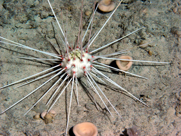
Adult Phoromosa placenta (Echinodermata: Echinoidea) at a depth of ca. 500 m from the Bahamas. |
Although each project requires different analytical capabilities, we have access to a variety of forms of microscopy: scanning laser confocal microscopy, scanning electron microscopy, fluorescence microscopy and light microscopy. |
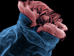
Colorize scanning electron micrograph of the anterior end of the rotifer (Brachionus plicatilis) showing the arrangement of the ciliated bands that compose the locomotory and feeding structure of rotifers, the corona. |
| Below are examples of representative images of developmental stages of different invertebrate animals that we have previously or are currently studying. | |
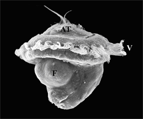
| This is a scanning electron micrograph of a larva of the Antarctic Limpet (snail) Nacella concinna. The specimen was reared and preserved at Palmer Station, Anvers Island, Antarctica. AT = apical ciliary tuft, F = developing foot, V = locomotory velum (note the hair-like cilia). |
|
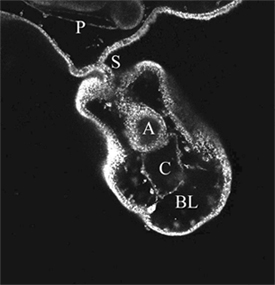
| This is an optical section through a developing clone of a sea star larva. The specimen was stained with Nile Red and the image was collected using a JEOL scanning laser confocal microscope. A = archenteron, BL = blastocoel of clone, C = clonal coelom, P = perivisceral coelom of the "parent" larva, S = stalk connecting parent larva to its offspring. |
|
|
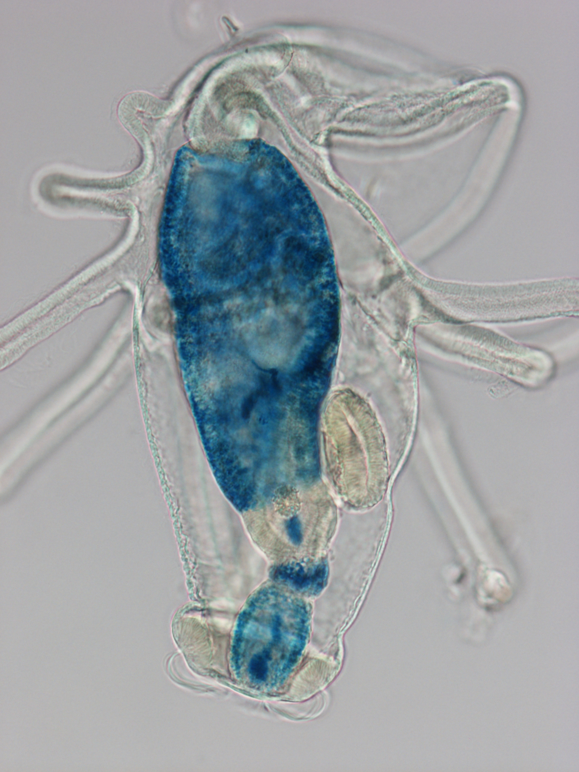
This is a right lateral view of an actinotroch larva (Phoronis architecta) collected at the Smithsonian Marine Station, FL in May 2012. The specimen was exposed to the iron-containing protein ferritin (0.5 mg / mL) for 4 hours. The presence of the iron (from ferritin) in the cells of the digestive system of this specimen was revealed through the "Prussian Blue" reaction. |
|
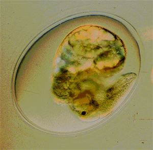
| This is not a picture of a larva. Rather this is a photograph of a developmental stage of a freshwater snail (Physa acuta). Members of this group have direct development and forego a larval stage. The shell, foot, and the dark eye are readily visible in this near term individual. It will eventually gnaw a hole on the capsule wall and hatch. |
|